Sierra Medical
Case Study
Ophthalmic Surgical Microscope VR Display Integration
Sierra Medical aimed to integrate a high-resolution VR display into its existing ophthalmic surgical microscope system. Using the Pimax Crystal Super 8K Micro-OLED, Pimax enabled direct DisplayPort video input and customized USB-based headset control, including wake-up and interpupillary distance adjustment. The solution allows Sierra Medical to retain its existing image acquisition system while managing VR display functions through its own equipment, without requiring a separate computer.
Healthcare / Medical Devices
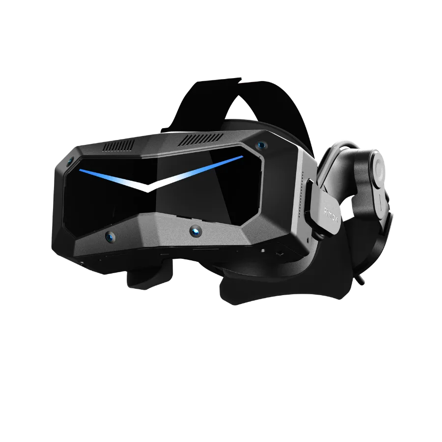 Pimax Crystal Super 8K Micro-OLED
Ophthalmic Microscope VR Display Integration
As ophthalmic surgical equipment becomes more digital, higher in resolution, and more integrated, medical device manufacturers are placing greater demands on microscope imaging displays. Beyond image clarity and real-time performance, key selection criteria include whether a display can connect directly to an existing image acquisition system and be managed by the equipment’s main controller.
Sierra Medical sought to improve the quality of its ophthalmic microscope imaging while integrating a high-resolution head-mounted display into its existing surgical microscope system. Using the Crystal Super 8K Micro-OLED as the display terminal, Pimax adapted microscope image display, headset wake-up, and interpupillary distance adjustment through a direct DisplayPort video connection and a customized USB control protocol. This enabled the customer to retain its existing image acquisition system and manage headset display and control through its own equipment.
The project has completed system integration testing and received customer acceptance.
Customer
Sierra Medical
Founded in July 2022 and based in Suzhou, China, Sierra Medical develops, manufactures, and sells equipment and associated consumables for minimally invasive ophthalmic surgery. Its products address areas including cataract and vitreoretinal treatment, and include ophthalmic surgical platforms, phacoemulsification equipment, vitreous cutters, intraocular laser fibers, and ophthalmic trocars.
As digital surgical visualization advances, medical device manufacturers are exploring alternatives to traditional optical eyepieces and external monitors. High-resolution head-mounted displays offer another way to present microscope images. Their practical use, however, depends on more than screen specifications: video interfaces, display latency, and compatibility with equipment control systems also matter.
Client Challenges
Upgrading the Display While Integrating with Existing Equipment
Before this collaboration, Sierra Medical had already incorporated a binocular 4K Micro-OLED head-mounted display into its development setup and wanted to further improve resolution, pixel density, and overall image quality. Upgrading the display terminal, however, involved more than replacing a VR headset with a higher-resolution model.
Sierra Medical already had an independent microscope image acquisition and processing system. The new display needed to receive video from that equipment and be managed by the customer’s own control system, without requiring an additional conventional PC VR application.
The two teams therefore needed to address three questions:
How could microscope images be delivered to the headset?
Optical images from the microscope needed to be converted into digital video by high-definition cameras and an acquisition board, then transmitted to the headset’s display system. The integration needed to ensure correct video input and complete images, while avoiding unintended cropping, missing image content, or obvious color shifts.
How could real-time display requirements be met?
Ophthalmic microscope imaging places demanding requirements on real-time video transmission. The project specified image latency below 20 ms as one of its technical requirements. Reducing the conventional PC VR application rendering stages helped streamline the path from image acquisition equipment to the display terminal.
How could the medical equipment control the headset?
In professional equipment integration, the headset must support the customer’s operating workflow as well as display images. Sierra Medical wanted its equipment’s host controller to send commands for functions such as headset wake-up and interpupillary distance (IPD) adjustment. The project specified an IPD adjustment range of 58–72 mm.
Pimax therefore needed to customize the device control protocol so that the headset could work with the customer’s existing control system.
Pimax Solution
High-Resolution VR Display Integration with Customized Device Control
To address these requirements, Pimax used the Crystal Super 8K Micro-OLED headset as the basis for adapting video display and device control to Sierra Medical’s existing microscope system. Microscope images continued to be generated by the customer’s acquisition and processing equipment, while the Pimax headset primarily served as a high-resolution near-eye display terminal.
Hardware
Pimax Crystal Super 8K Micro-OLED
High-Resolution Micro-OLED Display
The project uses a Crystal Super 8K Micro-OLED display configuration with the following key specifications:
| Parameter | Project Configuration |
|---|---|
| Display technology | Micro-OLED |
| Resolution per eye | 3840 × 3552 |
| Total pixels across both eyes | Approximately 27.3 million |
| Angular pixel density | 53 PPD |
| Video transmission | DisplayPort 1.4 |
| External control | Customized USB protocol |
Micro-OLED’s high pixel density provides a hardware foundation for presenting fine image detail. In medical imaging systems, however, final image quality also depends on the original image acquisition quality, video signal, optical system, and display settings.
The focus of this project was therefore to integrate high-resolution display capabilities into the customer’s existing microscope imaging system, rather than simply increase headset screen specifications.
Software & Integration
Direct Video Integration & Customized Headset Control
Direct DisplayPort Video Connection: Integrating with the Existing Microscope Imaging System
In conventional PC VR use, a headset typically relies on a computer to run a VR application and generate images. Sierra Medical’s microscope system already had independent image acquisition and processing capabilities, so an additional VR application was not needed to generate microscope images.
Pimax adapted the DisplayPort video interface so that video generated by the customer’s equipment could be sent directly to the headset’s display system.
The solution does not require a computer to render a PC VR application or run separate headset control software, while retaining the customer’s original image acquisition and processing architecture.
The customer’s equipment acquires and processes microscope images, then sends them directly to the headset through the adapted video interface. As a professional imaging display terminal, the headset can display the images for this project without connecting to a computer.
Customized USB Control Protocol: Enabling the Customer’s Equipment to Manage the Headset
In addition to video display, Sierra Medical wanted its own equipment control system to manage the headset’s essential functions without relying on separate PC-based headset control software. Pimax supported software customization and firmware updates so that the customer’s host controller could send commands to the headset through a USB control protocol and read the corresponding response messages.
The control functions involved in the project include:
Headset wake-up
Managing the headset’s operating state through external control commands.
IPD adjustment
Performing the relevant adjustment operations through the protocol according to the customer’s equipment requirements.
Command feedback
Returning corresponding messages for the customer’s system to check command execution status.
By adapting video display and device control separately, the headset can operate within the customer-defined workflow rather than function solely as a standalone consumer VR peripheral. This is one of the central benefits of the project’s custom development.
System Architecture
System Architecture: Microscope Image Transmission and Headset Integration
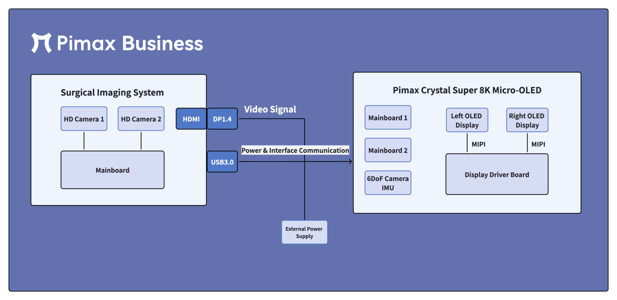
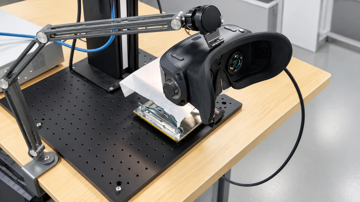
The system comprises microscope image acquisition equipment, external power and interface communication modules, and a Pimax head-mounted display terminal. Two high-definition cameras capture the microscope images. After processing by the acquisition board and video interface components, the images are transmitted to the headset through a DisplayPort 1.4 connection. Inside the headset, the display driver board connects to the left and right Micro-OLED screens through MIPI interfaces to present the images. In parallel, the customer’s equipment can manage functions such as headset wake-up and IPD adjustment through the project-specific USB control protocol.
The architecture handles video transmission and device control separately, allowing a high-resolution headset to be integrated as a display component within a professional medical imaging system.
Implementation & Outcomes
From Technical Evaluation to Customer Acceptance
Unlike standardized VR hardware procurement, professional medical equipment integration requires requirements confirmation, technical evaluation, and system integration testing using the customer’s actual equipment.
The project progressed through six main stages:
- 01
Requirements & Scenario Confirmation
Through requirements discussions and in-person exchanges, the teams clarified headset selection for a multi-user setting, the source of microscope images, how the headset would be used, and the customer’s equipment control requirements.
- 02
Technical Capability Evaluation
The teams assessed the Crystal Super’s display specifications, video interfaces, and control capabilities to identify what the existing hardware could support and which functions required custom development.
- 03
System Architecture Design
The teams confirmed the connections among image acquisition equipment, video interfaces, power and communication modules, and the headset, and defined the video transmission and device control implementation.
- 04
Imaging Performance Validation
Using the actual equipment, the teams checked video input, focusing on whether image completeness, cropping, color reproduction, and distortion met project requirements.
- 05
Software Customization & Protocol Integration
Software customization and firmware updates were accompanied by integration testing of USB control commands, response messages, and device behavior, enabling the customer’s system to invoke functions in the agreed manner.
- 06
Customer Acceptance
The customer validated microscope image display, IPD adjustment, and other project requirements, and completed project acceptance.
Project Outcomes
Through this development and validation work, the teams completed three areas of system adaptation:
Microscope image input
Established a DisplayPort video transmission path from the customer’s image acquisition equipment to the Pimax headset and completed image display validation.
Integrated device control
Implemented the headset wake-up, IPD adjustment, and command feedback functions required for the project through USB protocol customization.
Compatibility with the existing system
Retained the customer’s original image acquisition and processing architecture, reducing reliance on conventional PC VR application rendering and separate headset control software.
This process illustrates the difference between professional equipment integration and ordinary hardware procurement: delivery requires interface adaptation, protocol development, and customer-side validation in addition to the display hardware itself.
Potential Applications
From Ophthalmic Microscopes to Other Medical Imaging Equipment
Although this project focused on an ophthalmic surgical microscope, its approach to video interface adaptation and external control protocol customization can also inform other professional medical imaging applications.
Endoscopic and Laparoscopic Imaging Systems
For equipment with digital video outputs, it may be feasible to integrate a head-mounted display into the existing imaging path, subject to evaluation.
OCT and Other Medical Imaging Equipment
A high-resolution near-eye display solution can be evaluated against the specific image formats, output interfaces, and display requirements. Functions such as multi-source image fusion would still require additional software development.
Surgical Robot Operator Stations
These systems also involve video transmission, display latency, and control interfaces, but require fresh technical and safety assessments for each specific system.
These applications share certain technical characteristics with this project. This does not mean that the solution has already been validated with those types of equipment.
Frequently Asked Questions
Frequently Asked Questions
Can a VR Headset Connect Directly to a Surgical Microscope?
This requires an assessment of the microscope’s video output interfaces and signal formats, together with the headset’s underlying capabilities.
In this project, Pimax integrated the microscope imaging system with the headset through DisplayPort video input and custom development. This does not mean that every standard VR headset can connect directly to any microscope.
Does the Direct DisplayPort Connection Eliminate the Need for a Computer?
Yes. In this project’s direct DisplayPort video solution, microscope images can be sent directly to the headset for display without connecting to a computer.
After the microscope system acquires and processes the images, it sends them to the headset through the adapted video interface. No computer is needed to render a PC VR application or run separate headset control software.
This capability is based on the project’s interface adaptation and custom integration. It does not mean that all standard VR headsets support direct connections to arbitrary microscopes.
Does Pimax Support Custom Development for Medical Equipment?
Pimax can assess video interface adaptation, device control protocols, and related software or firmware customization according to the needs of a specific project.
This case demonstrates the use of DisplayPort video input and USB control protocol customization in an actual medical equipment integration project.
Can This Integration Approach Be Applied to Other Medical Imaging Equipment?
For endoscopes, OCT systems, and other professional equipment with digital video outputs, the project’s separation of video display and device control can serve as a reference. However, interfaces, image formats, latency requirements, and safety requirements vary across systems, so a new technical evaluation is necessary.
Professional Equipment Integration
From High-Resolution Displays to Professional Equipment Integration
For medical device manufacturers, selecting a head-mounted display involves more than comparing resolution, pixel density, and display technology. It is also essential to evaluate compatibility with existing video outputs, control protocols, and operating workflows.
In Sierra Medical’s ophthalmic surgical microscope project, Pimax used the Crystal Super 8K Micro-OLED as the display platform and integrated microscope image display and device control through DisplayPort video input and USB control protocol customization, receiving customer acceptance.
The case illustrates Pimax’s ability to support professional equipment manufacturers with interface adaptation, software customization, and system integration testing in addition to high-resolution display hardware.
Evaluate Your Medical Equipment Display Integration Requirements
If your company is developing ophthalmic surgical microscopes, medical imaging systems, or other professional equipment and requires high-resolution head-mounted displays, video interface adaptation, or device control protocol customization, contact the Pimax Business team at inquiry@pimax.com to discuss your technical requirements.

